Authors: V. Natesh*, Sangita Bhandary**, Shyam Thapa Chettri***, Bajrang Prasad****, Siddarth Koirala*****
* Assistant Professor of ENT & HNS, ** Professor and Head of ENT & HNS, *** Associate Professor of ENT & HNS, **** Senior Resident of ENT & HNS, ***** Associate Professor of Anesthesiology.
The authors declare that they have no financial disclosures or conflict of interest.
Institution:
Dept. of ENT and HNS, B P Koirala institute of Health Sciences, Dharan, Nepal
Corresponding Author:
DR V. Natesh
Assistant Professor, Department of ENT
B. P. Koirala Instituts of Health Sciences
Dharan, Nepal
E-mail: This email address is being protected from spambots. You need JavaScript enabled to view it.
Abstract:
Purpose: Foreign body aspiration remains a major cause of morbidity in children without early treatment. Management of aspiration requires a multidisciplinary approach and innovative approaches have helped reduce complications of bronchoscopy. We present a prospective and analytical review of clinical evaluation and management of cases with diagnosis or clinical suspicion of tracheobronchial foreign body aspiration. This study was done in a tertiary care teaching hospital in eastern Nepal, from August 2010 to December 2011.
Experimental Design: 12 cases with confirmed diagnosis or clinical suspicion of tracheobronchial foreign body aspiration that were admitted to our center between August 2010 and December 2011 underwent rigid bronchoscopy and telescopic removal of foreign body as per departmental protocol for management of airway foreign bodies. Successful retrieval of foreign body, its type, procedural events and post procedure events were analyzed.
Findings and Significance: All 12 cases which underwent rigid bronchoscopy under general anesthesia had a positive bronchoscopy. Male children comprised the majority of the study subjects and the average age of the series was 3.8 years. Peanut was the most common aspirated foreign body and the right main bronchus was the most common location of aspiration.
Summary & Conclusion: Foreign body aspiration (FBA) is a common cause for respiratory emergency in young children. An accurate history and careful examination aided by radiological findings are helpful in the diagnosis of FBA. Retrieval of the airway foreign body involves coordinated teamwork. Parental education and appropriate supervision of children during feeding is necessary for reducing the incidence of FBA.
Introduction:
Aspiration of foreign body into the respiratory tract, its varied clinical presentation, proper diagnosis and management has posed challenging questions for both otorhinolaryngologists as well as pediatricians for a long time. Foreign body aspiration (FBA) is a respiratory emergency and must be mannaged immediately or there can be a fatal outcome. Although there has been a decrease in complications associated with airway aspirations because of the significant advances in airway management and endoscopic technology, the incidence of FBA has not changed significantly.1 Literature regarding this entity is still scarce, more so from the south Asian region.
FBA is a serious health problem in pediatric patients causing significant morbidity and mortality.2 The need for improved knowledge led to the development of the Susy Safe Project, which maintains a data base of injurys caused by foreign body injestions.3 The leading cause of death in children less than three year of age is foreign body aspiration (FBA). The medical literature lists inorganic orbjects such as plastic and metal parts of tois as the most commonly aspirated objects.
Till the time of writing this article the total number of submitted FB cases in the Susy Safe Project was 17644, with maximum cases being less than 3 years of age. Similar data from USA shows that 500 children per year lose their life due to foreign body aspiration.4 For infants up to 12 months old, the aspiration of a foreign body is responsible for 40% of accident-related deaths. Usually small parts of food or other objects are aspirated.5 Approximately 10% of patients are infants.6 Delayed or poorly developed molars, along with poor meuromuscular development and coordiantion are risk factors. In addition, habitual placement of objects in the oral cavity for oral exploration are some important attributable causes for FBA in young children.7 A high index of suspicion and timely intervention can reduce morbidity as well as mortality especially in the pediatric age group. Time lag between the accident and expert attention is very important with regard to overall morbidity and mortality in a case of FBA.
Our aim was to study the clinical presentation, management and eventual outcome of foreign body aspiration (FBA) in children presenting to a tertiary care referral and teaching hospital, the only one in the eastern part of Nepal.
Methods:
A review and analysis of 12 cases with clinical suspicion/diagnosis of tracheobronchial foreign body aspiration, which underwent rigid bronchoscopy between August 2010 and December 2011 is presented. The subjects were taken for bronchoscopy on the basis of a confirmed diagnosis or strong clinical suspicion of FBA.
All cases underwent bronchoscopy as a planned procedure in the routine operating hours under general anesthesia using a Karl Storz (Germany), rigid ventilating bronchoscope with distal cold light illumination. Patients were put in a supine position with a blanket under the head to extend the atlanto-occipital joint and to align the oral, pharyngeal and tracheal axis in the same plane. Bronchoscope was introduced into the airway with the assistance of a Miller or Macintosh laryngoscope after the patient was well ventilated with a mask. The anesthetic T-piece was connected to the bronchoscope for ventilation and anesthesia. The rigid bronchoscope size for children was selected based on their age and expected cricoid diameter as per the table below.8
| Age | Cricoid Airway Diameter (mm) | Size | Bronchoscope Size | ED (mm) |
| 1 yr | 5.5 | 3.5 | 4.9 | 5.7 |
| 2 yr | 6.5 | 3.5 | 4.9 | 5.7 |
| 3 yr | 7.0 | 4.0 | 5.9 | 6.7 |
| 5 yr | 8.0 | 5.0 | 7.0 | 7.8 |
In most cases bronchoscope of 3.5 mm internal diameter and 20 cm length was used. The foreign bodies (FBs) were retrieved under 00 rigid telescopic view with a grasping optical peanut forceps in all cases after optimal suctioning around the FB. All the cases received antibiotic (Amoxycillin–Clavulinic acid) for 7 days and systemic steroids (Dexamethasone, 0.1 mg/kg 12 hourly) for 48 hours after the procedure. The subjects also received chest physiotherapy by a trained physiotherapist till the time of their stay in the ward. After bronchoscopy, all patients were NPO and received supplemental oxygen. Their heart rate, respiratory rate and oxygen saturation were closely monitored. Oral feeding was started once all vital parameters were normal. Post procedure chest X-rays were not done routinely and considered only in cases not showing anticipated resolution of symptoms. Subjects were discharged when vitals were stable, presenting symptoms had resolved and physical examination did not show any abnormality.
Demographic details (age and sex), history and duration of symptoms, physical examination, radiographic findings, bronchoscopic findings (nature of foreign body, its location, number of attempts for retrieval and duration of procedure), post bronchoscopy course, complications and outcome were analyzed.
Results:
12 subjects underwent bronchoscopy for confirmed/suspected foreign body aspiration at our center during a period of 17 months. 9 (75%) were males and 3 (25%) females. The mean age of subjects was 3.8 years; the youngest child being an 11 month old male child who had aspirated pieces of peanut and the oldest a male child of 7 years who had a board pin removed on bronchoscopy.
One subject had undergone bronchoscopy elsewhere prior to referral to our center as the endoscopist failed to retrieve the FB. Six (58%) children had a definite history of aspiration, whereas four children had prolonged respiratory symptoms or a history of recurrent pneumonia with doubtful or negative history of aspiration and were referred by our pediatric colleagues for bronchoscopy; a definitive FB was retrieved in 3 of these cases.
The minimum duration of symptoms was 1 day and maximum duration was 14 days. Overall, average duration of symptoms was 5.1 days. Cough and respiratory distress of varying degree were the predominant symptoms. Depending upon the type of foreign body, duration and site of lodgement; respiratory system examination revealed conducted sounds, decreased air flow, wheezing and crepitation.
Chest radiographs were done in all 12 cases. Unilateral hyperinflation (Figure 1) was the common finding seen in six (50%) of the cases, unilateral pneumonic consolidation (Figure 2) was seen in four (30%) cases, while in one case atelectesis was seen on X-ray. The 11 month child with peanut aspiration of one day duration had a normal X-ray. One patient with a diagnosis of Laryngotracheobronchitis (LTR) was on mechanical ventilation prior to bronchoscopy and showed bilateral consolidation. The child who had undergone a failed bronchoscopy elsewhere and was transferred to our center as the suspected foreign body could not be retrieved, showed consolidation in the radiograph.
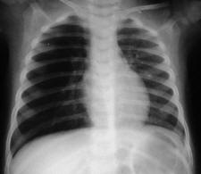
Figure 1. Hyperinflation right side on X-ray of chest in case no. 10 (Click on Picture To Enlarge)

Figure 2. Consolidation on right side on X-ray of chest in case no. 8

Figure 3. Appearance of Right Mainstem Foreign Body on Chest X Ray in case no. 10 (Click on Picture To Enlarge)


Figure 4. Rigid ventilating bronchoscopes of different sizes (2.5 to 4.5) and Oo telescopes with optical forceps for foreign body removal.
The majority of the FBs was of vegetable origin. Peanut (Figure 5) was the predominant foreign body being retrieved in eight cases (66.6%). In two cases, the foreign body was plastic in nature whereas in one case the foreign body had both a plastic and metal component (Figure 6). In another case referred for bronchoscopy, the cause of airway obstruction was thick necrotic mucous plug mainly in the right main bronchus and distal bronchi. In all our cases the foreign body was lodged either in the right main bronchus or the distal bronchus. The mean bronchoscope insertion per procedure was 3.5, which also included a mandatory insertion for diagnostic bronchoscopy. The maximum number of insertions was five and the minimum was two. In most of the cases, the total duration of the procedure was less than 20 minutes, whereas in 2 cases it was 24 minutes and 33 minutes, respectively.
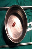

Figure 5. Peanut retrieved in total from the right main bronchus in case no. 10. Peanut pieces retrieved from the right main bronchus and distal bronchi in case no 3.


Figure 6. A plastic whistle retrieved in total from the right main bronchus in case no. 6. A board pin retrieved from the right main bronchus in case no. 12.
Three patients had post bronchoscopy bronchospasm that was managed in the Pediatric Intensive Care Unit (PICU). One child (case 11) required mechanical ventilation after bronchoscopy but was successfully weaned off the ventilator after 8 days and a second bronchoscopic clearance of mucous and necrotic tissue. The subject who underwent repeat bronchoscopy 2 days after the first for a failed bronchoscopy elsewhere, had pieces of peanuts removed from a sub-segmental bronchus and she recovered thereafter. There was no mortality in our series. The mean hospital stay was 3.9 days, the maximum duration of stay being 12 days and minimum being 3 days. All subjects were followed up biweekly for 2 weeks after discharge and did not report any residual or related new symptom.
Discussion:
Aspiration of foreign body into the tracheobronchial tree is not an uncommon problem in the pediatric population and the management of every case is a challenging clinical exercise. A variety of foreign bodies may be aspirated and may present in different ways to pose a diagnostic dilemma. The severity of clinical features depends upon the age of the patient, size, site, and type of foreign body aspirated as well as the duration of its lodgment. Usually a foreign body in the airway is common in the 1-3 years of age group.9 Children between 1 and 3 years of age have a habit of orally scrutinizing an object, don’t masticate well, have improper control of deglutition and are ambulatory during meals, all of which can predispose to aspiration.10 In our study, the average age of subjects was 3.8 years, the youngest child being 11 months old. In similarity to other studies, our series had a male preponderance (3:1) a fact attributed to their high activity.11
Diagnosis of FBA is often delayed because symptoms and signs are attributed to other causes of respiratory distress. In our series the mean duration of symptoms was 5.1 days, the maximum duration being 14 days. We believe that delayed presentation is a risk factor for post bronchoscopy complications and could be attributed to poor awareness among parents. Three parameters including history of aspiration, physical examination and a plain chest radiograph are very helpful diagnostic aids. Appropriate history and strong clinical suspicion are very informative and helpful for diagnosis but a negative history may be present in about 15% cases of foreign body inhalation.12 Even in our study, we found that half of the children had a definitive history of aspiration and choking witnessed by parents, whereas 33% of the children did not have a history of aspiration and a positive bronchoscopy was the result of a strong clinical suspicion of FBA.
96% to 97% of patients with FBA have clinical features of wheezing, caughing and unilateral decreased breath sounds, either alone or in combination.13 However, all children with the symptomatic triad of cough, respiratory distress and stridor had FBA (positive predictive value 100%). In our series, cough with reduced air entry in the effected side was the predominant clinical finding and correlated well with presence of FBA which is in accordance with other similar studies. Indeed, a combination of history, clinical signs and radiology is certainly more conclusive than any of them in isolation.
The Hallmark radiographic signs associated with foreign body aspiration are best demonstrated on expiratory plain chest X-ray findings.14 Fernandez, et al.15 emphasized that a 90% sensitivity in diagnosica can be achived with physical examination along with with respiratory auscultation. Chest radiography may reveal a variety of findings like unilateral air trapping, atelectesis, secondary pneumonic consolidation, and inspiratory obstruction or a combination of the findings may also be noted. As far as our findings in this regard is concerned, we found that a chest X-ray was an important tool in decision making for proceeding with a bronchoscopy and hyperinflation or consolidation were important findings in cases with FBA. The absence of positive radiological findings however does not exclude the diagnosis of foreign body aspiration as was true in one of our case. Diagnosis of radio opaque foreign body is easy when the whole respiratory tract is depicted but nonopaque foreign bodies need a cautious approach. The first step should be plain films in inspiration and expiration, and if needed fluoroscopy and CT scan should be done. Computed tomography is a useful noninvasive technique for guiding diagnosis and assessing the need for bronchoscopy, but is not recommended as a routine in the diagnosis of tracheobronchial foreign bodies. It is of value in difficult cases, in cases with radiolucent or soft-density foreign bodies, and is useful in defining complex anatomical areas.16 3D virtual bronchoscopy is a helpful aid in providing real time minute details of the whole tracheal-bronchial tree which optimizes the management of cases with FBA.
As much as FBA is a diagnostic challenge for both otorhinolaryngologist and pediatrician, rigid Bronchoscopy requires active coordination of both otorhinolaryngologist and anesthesiologist. The Storz ventilating bronchoscope is comprised of a metal tube with fibreroptic illumination and with air holes near the tip for ventilation of the contra-lateral lung. The distal end of the instrument also has a port for attaching the anesthetic Jackson Rees T-piece, a suction channel and a light prism. Selection of the appropriate size scope suitable for the patient’s airway is of utmost importance as it dictates the ease of ventilation and suctioning. A pertinent point to be kept in mind is that the nominal internal diameter (ID) is smaller than the actual diameter.
During bronchoscopy in response to compromised ventilation and gas exchange there is increase in sympathetic activity, decline in PO2, a rise in PCO2 and increase in airway resistance. At our center, general anesthesia is most commonly employed. Complications associated with anesthesia are hypoxemia, hypoventilation, hypertension, hypotension, tachycardia, myocardial ischemia, arrhythmias related to hypoxia and hypercarbia. Successful retrieval of the FB requires skilled coordination between the otorhinolaryngologist and anesthetist under optimal conditions.
Nuts and seeds are commonly aspirated by children. Peanut foreign bodys are the most common irrespective of the age of the child.17 In our series, peanuts were the most commonly aspirated object, accounting for 66% of the retrieved FBs. The types of inhaled foreign bodies will vary because of differing cultural practices, parental attitudes and eating habits. Young children with immature teeth are especially at risk for aspirating fragments of the nut because they cannot easily chew the peanut. The nut breaks into pieces and bronchoscopic removal sometimes becomes very difficult frequently requiring more than one attempt for complete removal. In our study, pieces of peanuts (Figure 5) were difficult to remove and required multiple attempts increasing the probability of complications. Accidental inhalation of foreign bodies is most commonly bronchial and the right side is more commonly involved as compared to the left.18 In our series, all the foreign bodies were retrieved from the right main bronchus and segmental bronchi. This can be attributed to the fact that the right main bronchus is relatively straightly aligned to the trachea and has a wider lumen.
Organic fragments cause greater inflammation than pieces of plastic or metal. Children inhaling nuts present with more severe symptoms and signs of respiratory distress, requiring urgent treatment. This is because the nut swells gradually and the oil and salt in the nut irritate the bronchial mucosa leading to an intense, local, chemical inflammatory reaction around the nut, making retrieval a difficult procedure. Vegetative foreign bodies swell up and get impacted in the bronchial wall producing ball valve obstruction, trapping air and leading to local emphysema or atelectesis, suppurative pneumonia, or bronchiectasis. Even if the object is removed, the inflammatory changes may take time to reverse, thus making post procedure management equally challenging.
Complications arising from bronchoscopy include post-operative pyrexia, atelectesis, pneumothorax, severe hemorrhage, laceration of bronchial wall, laryngospasm, bronchospasm and cardio respiratory arrest due to hypoxia and vagal stimulation. Late complications, especially with retained FB include formation of significant granulation, lung abscess and bronchiectasis.19 Retrieval of FB should only be attempted by an experienced team in a specialized center where there is a wide range of endoscopy equipment and appropriate anesthetic facilities available. Minimizing the number of attempts and total endoscopy time is essential in avoiding airway compromise. At our center we consciously attempt to limit the time of bronchoscopy to about 20 minutes and a maximum of three repeated insertions of the bronchoscope during one attempt to reduce post procedure complications. In our study, 3 children were managed postoperatively in the Pediatric Intensive Care Unit (PICU) for bronchospasm and inability to maintain SpO2 on room air after extubation. All these patients responded well to conservative management and were shifted out of PICU after 24 hours of observation, none of them requiring reintubation for ventilator support. One patient with a diagnosis of Laryngotracheobronchitis, required mechanical ventilatory support after bronchoscopic removal of a thick mucus plug and secretion with bronchoalveolar lavage. This child required one additional bronchoscopy for clearance of mucus and secretion and was successfully weaned off the ventilator after 8 days. We conclude that bronchospasm is the most common immediate post bronchoscopy complication and should be anticipated in every case more so in infants, foreign body of vegetable origin and subjects with coexisting airway infection.
The methods used in an emergency for retrieval of foreign body like the Heimlich maneuver, postural drainage or finger swiping the throat may worsen the outcome and should be discouraged.20 In any case with prolonged respiratory symptoms without clinical response to medical management, bronchoscopy is mandatory and should not be delayed irrespective of the age of the patient. A high index of clinical suspicion is mandatory for early diagnosis and management to prevent fatal outcome and long-term morbidity.
Conclusion:
FBA is a common and serious childhood emergency. The habit of oral exploration and access to small and inappropriate objects are contributory. Peanuts are the most commonly aspirated object and the right main bronchus and distal branch are the most common site of lodgment. History, clinical findings and radiology aid in diagnosis. Bronchoscopy under optimal conditions with a skilled surgical and anesthetic team is desirable.
References:
1. Tan HK, Brown K, McGill T, Kenna MA, Lund DP, Healy GB. Airway foreign bodies (FB): a 10-year review. Int J Pediatr Otorhinolaryngol. 2000 Dec 1;56(2):91-9. View Abstract
2. Dosios T, Safioleas M, Xipolitas N. Surgical treatment of esophageal perforation. Hepatogastroenterology. 2003 Jul-Aug;50(52):1037-40. View Abstract
3. Gregori D. The Susy Safe Project. A web-based registry of foreign bodies injuries in children. Int J Pediatr Otorhinolaryngol. 2006 Sep;70(9):1663-4. Epub 2006 Jul 10. View Abstract
4. Black RE, Choi KJ, Syme WC, Johnson DG, Matlak ME. Bronchoscopic removal of aspirated foreign bodies in children. Am J Surg. 1984 Dec;148(6):778-81. View Abstract
5. Reilly JS, Cook SP, Stool D, Rider G. Prevention and management of aerodigestive foreign body injuries in childhood. Pediatr Clin North Am. 1996 Dec;43(6):1403-11. View Abstract
6. 4. Black RE, Johnson DG, Matlak ME. Bronchoscopic removal of aspirated foreign bodies in children. J Pediatr Surg. 1994 May;29(5):682-4. View Abstract
7. Mu LC, Sun DQ, He P. Radiological diagnosis of aspirated foreign bodies in children: review of 343 cases. J Laryngol Otol. 1990 Oct;104(10):778-82. View Abstract
8. Roberts S, Thornington RE. Paediatric bronchoscopy. Continuing Education in
Anesthesia, Critical Care & Pain. 2005 5(2):41-44. Link to Article
9. Aytaç A, Yurdakul Y, Ikizler C, Olga R, Saylam A. Inhalation of foreign bodies in children. Report of 500 cases. J Thorac Cardiovasc Surg. 1977 Jul;74(1):145-51. VView Abstract
10. Daniilidis J, Symeonidis B, Triaridis K, Kouloulas A. Foreign body in the airways: a review of 90 cases. Arch Otolaryngol. 1977 Oct;103(10):570-3. View Abstract
11. Pyman C. Inhaled foreign bodies in childhood. (A review of 230 cases). J Otolaryngol Soc Aust. 1971 Mar;3(2):170-80. View Abstract
12. Kim IG, Brummitt WM, Humphry A, Siomra SW, Wallace WB. Foreign body in the airway: a review of 202 cases. Laryngoscope. 1973 Mar;83(3):347-54. View Abstract
13. Tomaske M, Gerber AC, Stocker S, Weiss M. Tracheobronchial foreign body aspiration in children - diagnostic value of symptoms and Swiss Med Wkly. 2006 Aug 19;136(33-34):533-8. View Abstract
14. JACKSON C. Bronchial obstruction. Dis Chest. 1950 Feb;17(2):125-50. View Abstract
15. Fernández Jiménez I, Gutiérrez Segura C, Alvarez Muñoz V, Peláez Mata D. [Foreign body aspiration in childhood. Review of 210 cases]. An Esp Pediatr. 2000 Oct;53(4):335-8. View Abstract
16. Alford BR, Chenault DI, Danziger J. Detection of foreign bodies with computerized tomography. Arch Otolaryngol. 1979 Apr;105(4):203-4. View Abstract
17. Fraga Ade M, Reis MC, Zambon MP, Toro IC, Ribeiro JD, Baracat EC. Foreign body aspiration in children: clinical aspects, radiological aspects and bronchoscopic treatment. J Bras Pneumol. 2008 Feb;34(2):74-82. View Abstract
18. Jackson, C, Jackson, CL. Diseases of the air and food passages of foreign body origin, WB Saunders, Philadelphia 1936. p.135.
19. Oliveira CF, Almeida JF, Troster EJ, Vaz FA. Complications of tracheobronchial foreign body aspiration in children: report of 5 cases and review of the literature. Rev Hosp Clin Fac Med Sao Paulo. 2002 May-Jun;57(3):108-11. View Abstract
20. Cohen SR, Herbert WI, Lewis GB Jr, Geller KA. Foreign bodies in the airway. Five-year retrospective study with special reference to management. Ann Otol Rhinol Laryngol. 1980 Sep-Oct;89(5 Pt 1):437-42. View Abstract